PLANNING AND TOPICS
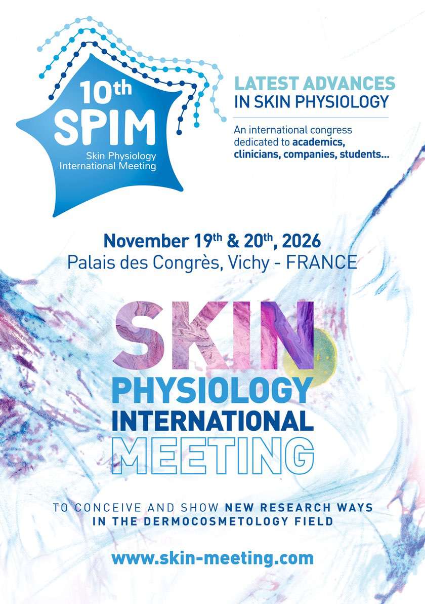
SPIM 2026 - 10thedition
NOVEMBER 19th
> LONGEVITY & HEALTHY SKIN
9.00 – 10.45 – Chairman: Prof. Jérôme LAMARTINE
-
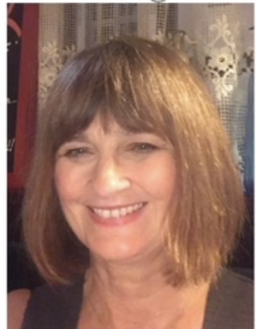
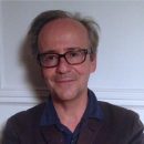
Dr. Sibylle JAGER, L’OREAL, Paris, France
“Longevity – is much within us still a worm ?” (Nietzche) View more -
Dr. Isabelle ADER, RESTORE Laboratory, INSERM 1301, IHU HealthAge, University of Toulouse, France
“Aging: skin cells as sentinels of healthy ageing” View more -
Dr. Maria CAVINATO, Institute for Biomedical Aging Research, University of Innsbruck, Austria
“Cellular Senescence in Skin Aging and Disease: Emerging Mechanisms and Translational Opportunities” View more
10.45 – 11.15 – COFFEE BREAK – POSTER SESSION
> JUNIOR RESEARCHERS
11.15 – 12.15 – Chairman: Prof. Frédéric CAUX
3 selected young scientists
12.30 – 14.00 – LUNCH BREAK
> MICROBIOMA & EPITHELIAL BARRIER
14.00 – 15.45 – Chairman: Dr. Lionel BRETON
-
Prof. Joël DORÉ, Paris-Saclay University, France
“Human-microbes symbiosis: A target for innovations in prevention and therapies” View more -
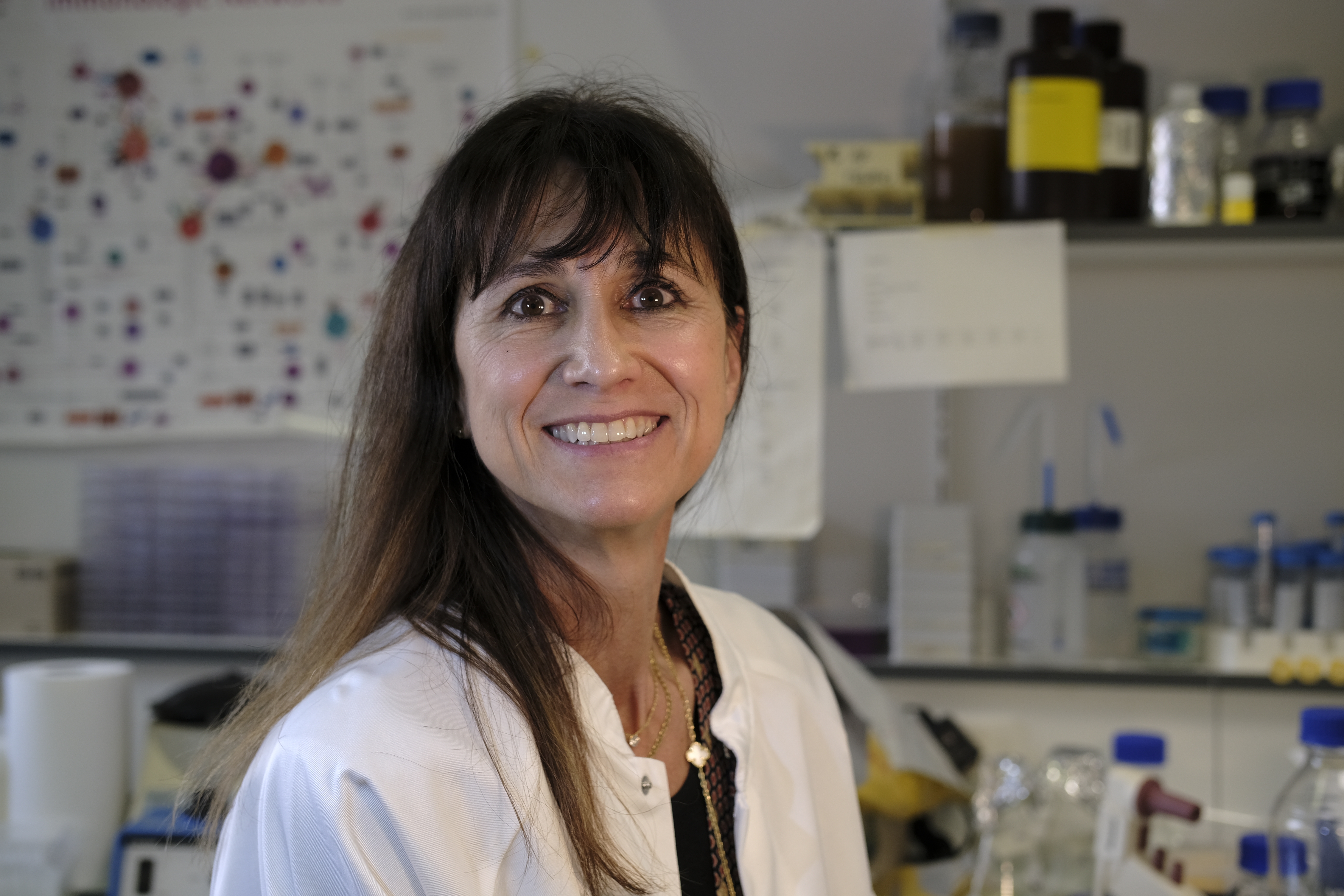
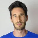
Prof. Julia OH, The Jackson Laboratory, Duke University School of Medicine, Durham, USA
“The human skin microbiome: microbial modulation of skin" View more -
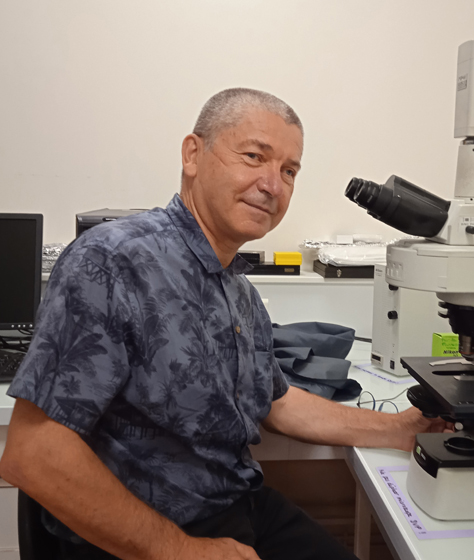
Prof. Marc G. FEUILLOLEY, University of Rouen Normandy, France
“Endocrine Microbiology, from gut to skin” View more
15.45 – 16.15 – COFFEE BREAK – POSTER SESSION
> JUNIOR RESEARCHERS
16.15 – 17.15 – Chairman: Dr. Corine BERTOLOTTO
3 selected young scientists
-

17.15 - 17.30 - Dr. Richard LEROUX, President of the SFC
Presentation of the French Society of Cosmetology (SFC) organisation -
17.30 - 19.00 Meeting with the SPIM 2026 painter
Judith GRASSL -
20.00 - Gala Dinner
NOVEMBER 20th
> LONGEVITY & HEALTHY SKIN
9.00 – 10.15 – Chairman: Dr. Maria DALKO-CSIBA
-
Dr. Anita KRISKO, University of Goettingen, Germany
“Proteostasis, respiration and metabolism: a ménage-a-trois of cellular longevity” View more -
Prof. Carmit LEVY, Faculty of Medicine, Tel-Aviv University, Israel
“Physiological benefit of UV exposure other than vitamin D production” View more
10.15 – 10.45 – COFFEE BREAK – POSTER SESSION
> MICROBIOMA & EPITHELIAL BARRIER
10.45 – 12.45 – Chairman: Dr. Marek HAFTEK
-
Prof. Ellen Van den BOGAARD, Radboud University, Nijmegen, Netherlands
“Various roles of AHR in epidermal Homeostasis and Host Defense” View more -
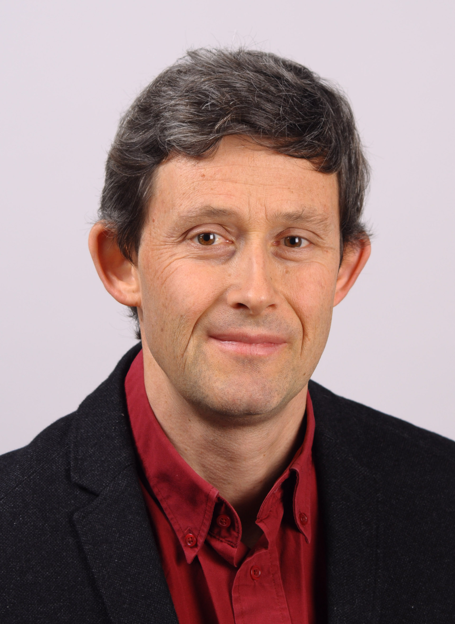
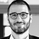
Dr. Cédric DELEVOYE, INSERM U1151/CNRS 8253, University of Paris Cité, France
“Genome photoprotection: connecting intracellular mechanics to pigment organelles
positioning" View more -
Dr. Patrick VEIGA, Institute MICALIS, INRAE, University of Paris-Saclay, Jouy-en-Josas, France
“From French gut to French skin“ View more
12.45 – 14.00 – LUNCH BREAK
> MICROBIOTA SESSION HOSTED BY THE FRENCH SOCIETY OF DERMATOLOGY (SFD)
14.15 – 16.15
-
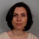
Prof. Saskia ORO, Henri-Mondor Hospital Créteil, Paris, France, President of the SFD
Introduction (5 min) -
Prof. Nicolas DUPIN, Cochin Hospital, Paris, France
“Cutibacterium acnes - Pathogenic potential and therapeutic Perspectives” (15 min) -
Dr. Julien SCANZI, Estaing Hospital, Clermont-Ferrand, France
Prof. Anthony BUISSON, Estaing Hospital, Clermont-Ferrand, France
“Gut microbiota: Understanding its implications in applied research” (15 min) -
Prof. Emilie SBIDIAN, AP-HP, Henri Mondor Hospital, INSERM U955, Créteil, France
“Impact of psoriasis biotherapies on the gut microbiota” (15 min) -
Dr. Claire HOTZ, Rodez Hospital, France
“Fecal Microbiota Transplantation in hidradenitis suppurativa: Why and how? “FESTIVAL”
project (15 min) -
Prof. Marie BEYLOT-BARRY, UMR 1312 INSERM/University of Bordeaux, France
Prof. Olivier CHOSIDOW, AP-HP, Bicêtre Paris-Saclay & Ambroise Paré-UVSQ, France
Q&A Round Table (50 min) -
Prof. Marie BEYLOT-BARRY, Bordeaux, France
Conclusion (5 min)
> GREENTECH AWARDS CEREMONY
16.15 – 17.00